UCSB Neuroscientists Study Connectivity in the Human Brain
The human brain is one of the most complex systems both in terms of its structural organization and of the diverse functionality that structure supports. One of the great challenges in modern neuroscience is to understand how human cognitive function arises from interactions between different regions of the brain.
Using magnetic resonance imaging (MRI) technology, researchers at UC Santa Barbara have identified organizational features of human brain anatomy that support coordinated changes in functional brain activity when an individual is at rest, attending to a visual task, or remembering something such as a word or face. Their work was highlighted in a recent issue of the Proceedings of the National Academy of Sciences.
"Identifying such relationships between anatomy and function is crucial for understanding how the anatomical organization of the human brain both supports and constrains human cognitive function," said lead author Ann M. Hermundstad. A postdoctoral researcher in the Department of Physics and Astronomy at the University of Pennsylvania, Hermundstad was a graduate student in the Department of Physics at UCSB when the research was conducted and the paper was written.
"There have been tens of thousands of experiments mapping out the cortex of the brain and showing the different areas associated with different activities," said Scott T. Grafton a professor in UCSB's Department of Psychological and Brain Science and director of the campus's Brain Imaging Center. "But it's a very modular view of the brain, and scientists have always known there's a lot more to it. There are networks involved, and sets of regions that work together."
Grafton and his team studied the relationship between various cognitive functions, such as attention and memory, and the associated brain networks. "There are nuances," Grafton explained. "And network activity depends on what you're doing. At rest, you see great relationships between the big bundles of wires that connect the two hemispheres, but you see really weak relationships within the hemispheres. The big bundles within the hemisphere don't really track with the activity that ought to be using them. As soon as you do some task, such as remembering a face, the networks within the hemispheres come into play and there is a strong correspondence between the strength of a wire and the strength of a signal." That makes sense, he noted, because so many human functions are specialized to one hemisphere or the other.
One surprising finding of the study, according to Hermundstad, is that the relationships between anatomy and function are observed globally across the entire brain. "We might have expected a more regionally specific relationship," she said. "This global relationship means that we can infer correlations in neural activity, which reflect the putative strength of communication between brain regions, by knowing only the properties of the anatomical pathways that link those brain regions."
This particular study lays the foundation for additional research in functional connectivity, as well as a host of other areas of study. "We can ask why, from a physical and biological standpoint, the brain is wired in this manner –– does the particular layout of anatomical pathways help facilitate efficient or reliable communication between many brain regions?" Hermundstad said. "We can similarly ask whether the relationship between brain anatomy and brain function impacts a person's ability to perform different tasks, such as the ability to learn new information or remember old information."
The findings also make specific predictions about how brain function might change if particular pathways change or become nonfunctional, Hermundstad noted. Such predictions are important for understanding many neurological disorders, such as schizophrenia, that are associated with altered brain anatomy.
Other UCSB researchers contributing to the study include Jean Carlson, professor of physics; Michael B. Miller, professor of psychology; Danielle S. Bassett, Sage Center for the Study of the Mind Junior Research Fellow in physics and in psychological and brain sciences; and Amy Frithsen and Arianne Johnson, graduate students in psychological and brain sciences. In addition, Kevin Brown, currently an assistant research professor of chemical engineering at the University of Connecticut, was a postdoctoral researcher in neuroscience at UCSB when the research was conducted.
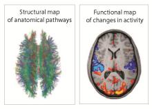
† Bottom image: MRI techniques enable the noninvasive measurement of structural pathways (white matter cables linking different brain regions, left) and functional pathways (coordinated changes in brain activity, right) in the human brain.
Credit: FMRIB Centre, University of Oxford
Related Links
Proceedings of the National Academy of Sciences
UC Santa Barbara Brain Imaging Center
Share this article
Related Stories