Seeing is Believing
A new, state-of-the-art facility dedicated to the finest imaging of soft and biological materials is in the works at UC Santa Barbara. Aided by a $1.5 million grant from the Arnold and Mabel Beckman Foundation, scientists will come closer to visualizing molecular-level structures, such as cellular components and polymer assemblies, in their native environments. The work will allow insights into relationships and functions, enabled by three-dimensional, cryogenic transmission electron microscopy (3D cryo-EM).
“We are launching a top-of-the-line facility at UC Santa Barbara from scratch,” said Songi Han, a professor in the Department of Chemistry and Biochemistry, who is spearheading the project. “I’m excited.”
The Arnold and Mabel Beckman Foundation Sample Preparation for Cellular Cryo-ET Award makes possible the acquisition of a Cryo-Focused Ion Beam (Cryo-FIB) Milling Instrument, a crucial piece of equipment for the soon-to-be UCSB Structural Biology cryo-Electron Microscopy (SB2EM) facility, to be located in the campus’s Chemistry Building. Combined with cryogenic fluorescence microscopy and sample preservation auxiliary equipment, the milling instrument will enable imaging of previously inaccessible samples and soft materials at increasing levels of spatial complexity.
“This is fantastic news,” said Pierre Wiltzius, dean of mathematical, life and physical sciences at UCSB. “Having the cryo-EM and the FIB will put us on a new growth curve.”
“We are extremely grateful for the support of the Arnold and Mabel Beckman Foundation,” said Tresa Pollock, interim dean of the College of Engineering and the Alcoa Distinguished Professor of Materials. “These major grants ensure that our faculty and students have access to the state-of-the-art facilities and equipment required to engineer solutions and to conduct leading-edge interdisciplinary research.”
Ice and Slice
Three-dimensional cryo-EM has been practiced as early as the 1970s, seen at first as an esoteric technology, then gradually moving closer to the mainstream. That’s according to Dorit Hanein, a professor at Scintillon Institute in San Diego with an adjunct appointment in UCSB’s Department of Chemistry and Biochemistry, and an expert in cellular tomography.
“It got a significant boost in the last 10 years because of significant advances in both instrumentation, and computational power,” Hanein said of innovations that would lead to the 2017 Nobel Prize in Chemistry, awarded to a trio of biophysicists and cryo-EM experts for a technology which both simplify, improve and provide the means to imaging of biomolecules in three-dimension in close to their native environment.
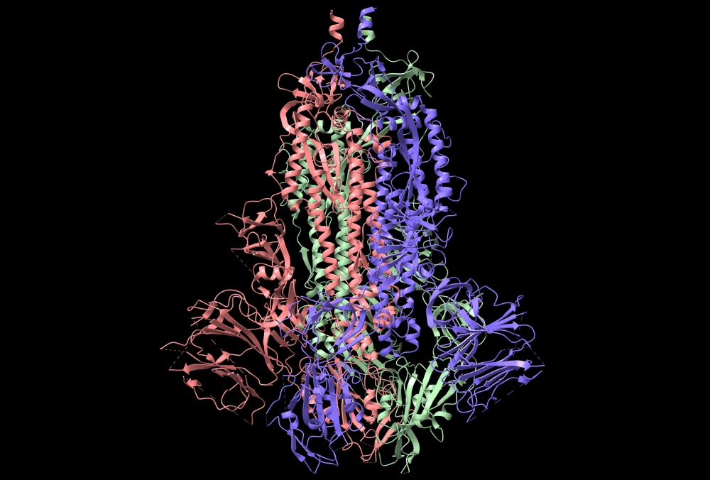
The sample preparation process involves rapid freezing of macromolecular assemblies, cells or tissue while in solution — freezing so fast that ice crystals can’t form, thus capturing the samples in their natural shapes while embedded in a glassy layer of frozen solution. To image the sample, a transmission electron microscope emits a beam of electrons that pass through the specimen, captured and aided by computational analysis that translates the data into three-dimensional representations.
“The range of information you can achieve is on various levels,” Hanein said, “from atomic information for single molecules and assemblies, seeing molecular details of the building blocks of cell and tissues while in their native environment.” Cryo-EM is able to capture specimens in more dynamic states that often reveal more than the traditional practice of isolating samples can, she added.
“This state-of-the-art capability puts a large community of researchers in the forefront of structural biology science and discovery and will have an impact on many related areas,” said Mattanjah DeVries, chair of chemistry and biochemistry.
Before the cryo-transmission electron microscope works its wonders, however, the sample must be prepared. More specifically, it has to be made very thin in order for the electron beams to pass through as needed.
“Making the samples for collecting the data is the main bottleneck,” Han said. For all the ability of the electron microscope to capture complexity, the samples still have to be ultrathin.
This is where the cryo-FIB milling instrument comes in. Its job is to carve out these ultrathin frozen sheets — called lamellae — in an automated process that can relieve the bottleneck for users of this facility.
“It’s a vital piece of the workflow that will allow us to look at those samples,” said Han, who anticipates that the new facility will be in demand, noting that several teams on campus are conducting research that could benefit greatly from the state-of-the-art equipment.
Among those investigators is Beth Pruitt, director of the campus’s Center for Bioengineering. “Cryo-EM microscopy with the FIB milling device will be transformative to research across campus,” she said. “I can’t wait to learn more about how sub-cellular proteins assemble to imbue structure and function to cells over long ranges.”
“This milling technique allows researchers to essentially open a window into the cell interior itself and see directly how all the components are interacting,” explained Beckman Foundation Executive Director Anne Hultgren. “An equally important part of these awards is in method development to couple fluorescence or another imaging method to the milling equipment so that the researchers can target these precise locations as they are preparing the samples for imaging in the high-resolution cryo-electron microscopes.”
The cryo-FIB milling instrument, combined with cryogenic fluorescence microscopy capabilities and sample preservation auxiliary equipment — all funded by the Beckman Foundation — and the transmission electron microscope (largely supported by shared instrumentation grant (S10) from the National Institutes of Health) will be installed in a suite custom-fitted for the equipment, climate controlled and isolated from electromagnetic interference and vibration. It’s a big project, the SB2EM facility that is expected to be completed by the end of 2022.
For Han, Hanein, Pruitt, colleagues Sid Dey (chemical engineering), Ryan Stowers and Niels Volkmann (mechanical engineering) and other collaborators it will be worth the wait.
“Many congratulations to Professor Songi Han and her collaborators for receiving this major grant from the Beckman Foundation to acquire a cryo-FIB,” said Rachel Segalman, chair of the Chemical Engineering Department and the Edward N. Kramer Professor of Materials. “This instrument, along with a cryo-transmission electron microscope that the university recently secured funding to purchase, represent a major leap for our campus in biomolecular engineering and structural biology research.”
Located in Irvine, California, the Arnold and Mabel Beckman Foundation supports researchers and nonprofit research institutions in making the next generation of breakthroughs in chemistry and the life sciences.
Share this article
Related Stories