Materials scientists working with biologists at the University of California, Santa Barbara have developed "smart" bio-nanotubes --- with open or closed ends --- that could be developed for drug or gene delivery applications.
The nanotubes are "smart" because in the future they could be designed to encapsulate and then open up to deliver a drug or gene in a particular location in the body. The scientists found that by manipulating the electrical charges of lipid bilayer membranes and microtubules from cells, they could create open or closed bio-nanotubes, or nanoscale capsules. The news is reported in an article to be published in the August 9 issue of the Proceedings of the National Academy of Sciences. It is currently available on-line in the PNAS Early Edition. See: http://www.pnas.org/cgi/content/abstract/0502183102v1
The findings resulted from a collaboration between the laboratories of Cyrus R. Safinya, professor of materials and physics and faculty member of the Molecular, Cellular, and Developmental Biology Department, and Leslie Wilson, professor of biochemistry in the Department of Molecular, Cellular and Developmental Biology and the Biomolecular Science and Engineering Program. The first author of the article is Uri Raviv, a post-doctoral researcher in Safinya's lab and a fellow of the International Human Frontier Science Program Organization. The other co-authors are Daniel J. Needleman, formerly Safinya's graduate student who is now a postdoctoral fellow at Harvard Medical School; Youli Li, researcher in the Materials Research Laboratory; and Herbert P. Miller, staff research associate in the Department of Molecular, Cellular and Developmental Biology.
The scientists used microtubules purified from the brain tissue of a cow for their experiments. Microtubules are nanometer-scale hollow cylinders derived from the cell cytoskeleton. In an organism, microtubules and their assembled structures are critical components in a broad range of cell functions –– from providing tracks for the transport of cargo to forming the spindle structure in cell division. Their functions include the transport of neurotransmitter precursors in neurons.
"In our paper, we report on a new paradigm for lipid self-assembly leading to nanotubule formation in mixed charged systems," said Safinya.
Raviv explained, "We looked at the interaction between microtubules –– negatively charged nanometer-scale hollow cylinders derived from cell cytoskeleton –– and cationic (positively charged) lipid membranes. We discovered that, under the right conditions, spontaneous lipid protein nanotubules will form."
They used the example of water beading up or coating a car, depending on whether or not the car has been waxed. Likewise the lipid will either bead up on the surface of the microtubule, or flatten out and coat the whole cylindrical surface of the microtubule, depending on the charge.
The new type of self-assembly arises because of an extreme mismatch between the charge densities of microtubules and cationic lipid, explained Raviv. "This is a novel finding in equilibrium self-assembly," he said.
The nanotubule consisting of a three-layer wall appears to be the way the system compensates for this charge density mismatch, according to the authors.
"Very interestingly, we have found that controlling the degree of overcharging of the lipid-protein nanotube enables us to switch between two states of nanotubes," said Safinya. "With either open ends (negative overcharged), or closed ends (positive overcharged with lipid caps), these nanotubes could form the basis for controlled chemical and drug encapsulation and release."
The inner space of the nanotube in these experiments measures about 16 nanometers in diameter. (A nanometer is a billionth of a meter.) The whole capsule is about 40 nanometers in diameter.
Raviv explained that the chemotherapy drug Taxol is one type of drug that could be delivered with these nanotubes. The scientists are already using Taxol in their experiments to stabilize and lengthen the lipid-protein nanotubes.
The work was performed using state-of-the-art synchrotron x-ray scattering techniques at the Stanford Synchrotron Radiation Laboratory (SSRL), combined with sophisticated electron microscopy at UCSB. The work was funded by the National Institutes of Health and the National Science Foundation. SSRL is supported by the U.S. Department of Energy. Raviv was also supported by the International Human Frontier Science Program and the European Molecular Biology Organization.
###
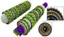
† About the Illustration
"Smart" bionanotubes. Lipid protein nanotubes made of microtuble protein (made of tubulin protein subunits shown as red-blue-yellow-green objects) that is coated by a lipid bilayer (drawn with yellow tails and green and white spherical heads) which in turn is coated by tubulin protein rings or spirals. By controlling the relative amount of lipid and protein it is possible to switch between two states of nanotubes with either open ends (shown in the center) or closed ends with lipid caps (shown on the left), a process which forms the basis for controlled chemical and drug encapsulation and release. A top view of the nanotubes and a magnified region is shown on the right. The image was created by Peter Allen.
Related Links
Share this article
Related Stories